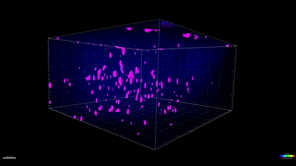
Two-photon imaging of Tau tangles in the brain of a living mouse. Data obtained at Neurotar.
We use in vivo two-photon microscopy to visualize the formation of Tau tangles (also known as neuro-fibrillary tangles or NFTs), their targeting by anti-Tau biologics, and the effects of small-molecule drugs on the tangles. This assay is optimal for preclinical studies of target engagement, mechanism of action, and Tau pharmacodynamics. The assay is also highly suitable for detailed mechanistic characterization of novel Tg mouse models of tauopathies.
Tau targeting strategies in Alzheimer’s research
Next to amyloid-beta (Aβ), tau proteins are key hallmarks of Alzheimer’s disease (AD) and targets of multiple disease-modifying therapies. Tau-targeting therapies are somewhat behind amyloid-targeted approaches in terms of clinical advancements. Nevertheless, several tau-directed therapies are making their way through clinical trials. The main focus is on antibodies that can attach to tau proteins and prevent their aggregation or facilitate their removal from the brain.
Similarly to Abeta-targeting interventions, tau-targeting therapies aim at mitigating cognitive decline by preventing aggregation or facilitating the clearance of tau protein tangles. Therefore, they stand to benefit from advanced imaging techniques such as MRI, PET, bioluminescence, and multiphoton imaging, also known as two-photon microscopy. Imaging approaches stand out for their ability to visualize the tangles and quantitatively measure tangle burden over time. They do so within the physiologically relevant context of the living brain.
Why study the Tau tangle dynamics with two-photon microscopy?
Tau tangles are micrometer-size intracellular structures and their visualization typically requires ex vivo microscopic analysis. Two-photon microscopy is uniquely suited for longitudinal analysis of tau tangle dynamics in vivo. This is due to its superb spatial resolution compared to other imaging techniques (Kuchibhotla et al., 2014; Wu et al., 2018).
Targeting Abeta instead of Tau? Follow this link to learn about our Amyloid beta dynamics imaging assay.
What types of studies can Neurotar perform for Tau-targeting drug developers?
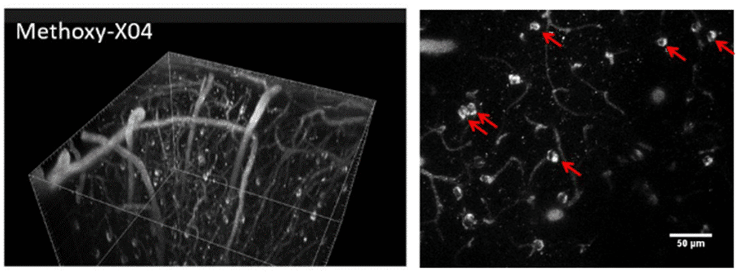
The study design starts with an assessment of your molecule’s size. It is possible to label the relatively large biological molecules with a fluorescent tag to add an extra pharmacokinetic/biodistribution dimension to a study. Measuring NFTs’ proximity to anatomical structures such as blood vessels requires additional labeling, for example with fluorescently labeled dextrans.
The most popular options for our clients’ Tau tangle dynamics imaging studies include:
We offer all of the standard administration routes, such as i.v., i.p., and per oral gavage. In addition, we can also perform intracranial or intrathecal injections of non-BBB-crossing drug candidates.
A popular extension for the NFT imaging studies is the addition of behavioral observations such as hind paw clasping score and weight loss.
We can also help you identify the NFT-expressing neuronal population. This can be achieved by overexpressing a fluorescent marker of neuronal morphology (e.g., YFP or RFP). To do this in certain neuronal populations, we typically use viral delivery and neuron-type-specific promoters.
How can Neurotar facilitate your in vivo studies on NFT-expressing mouse models?
We shall begin by learning from you about your research goals and designing an optimal NFT imaging study together.
Unless you already have a fluorescently labeled compound or biomolecule, we shall assist you in setting up a labeling protocol.
Together, we will select a suitable Tg mouse model such as rTG4510, Tau.P301L, or Tau.P301S. We can help you source these mice from one of our verified providers such as reMYND.
Neurotar will then implant the cranial windows one month before the first imaging session, run the experiment, collect and analyze the data, and send you a preliminary report along with our proposed interpretation of the study results. Afterward, we shall answer all of your questions and integrate your suggestions into a final report. If you’d like to publish the results, we’ll be happy to help you write the Materials and Methods section.
Cited works:
Kuchibhotla KV, Wegmann S, Kopeikina KJ, Hawkes J, Rudinskiy N, Andermann ML, Spires-Jones TL, Bacskai BJ, Hyman BT (2014). Neurofibrillary tangle-bearing neurons are functionally integrated in cortical circuits in vivo. PNAS. 111(1):510-4. https://doi.org/10.1073/pnas.1318807111
Wu Q., Lin Y., Gu J., Sigurdsson EM. (2018). Dynamic assessment of tau immunotherapies in the brains of live animals by two-photon imaging. eBioMedicine 35, 270-278. https://doi.org/10.1016/j.ebiom.2018.08.041
Other two-photon brain imaging services offered by Neurotar
| Neurodeg. Disease (e.g. AD, PD) | Stroke and TBI | Neuropathic Pain and Migraine | Neuropsychiatry (e.g. Schizophrenia) | Epilepsy | |
|---|---|---|---|---|---|
| Blood-brain barrier integrity | ✓ | ✓ | ✓ | ||
| Trans-BBB pharmacokinetics | ✓ | ✓ | ✓ | ||
| Dendritic spine turnover | ✓ | ✓ | ✓ | ✓ | ✓ |
| Microglial dynamics or response to injury | ✓ | ✓ | ✓ | ✓ | ✓ |
| Calcium signaling | ✓ | ✓ | ✓ | ✓ | ✓ |
| Abeta Plaque or Tau Tangle dynamics | ✓ | ✓ | ✓ | ||
| Mitochondrial fragmentation | ✓ | ✓ | ✓ | ✓ | ✓ |
| Ischemic Stroke model | ✓ | ||||
| Regeneration of peripheral neurons | ✓ | ✓ | ✓ |